
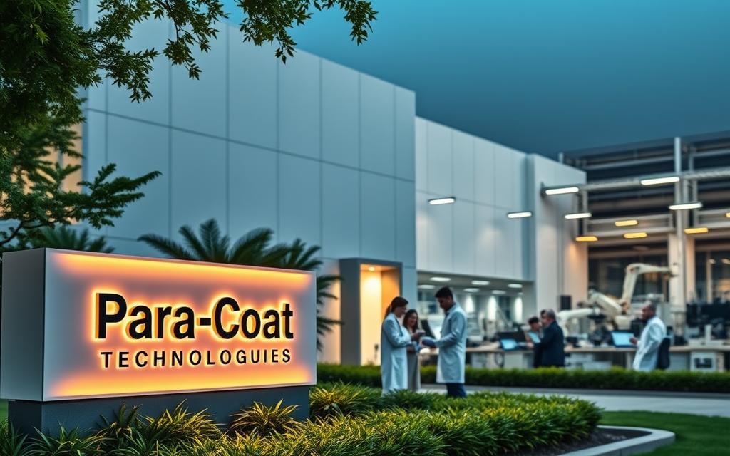
In the world of advanced manufacturing, coating services are key to protecting sensitive parts. Para-Coat Technologies Inc., based in Johnstown, Pennsylvania, is leading the way in this field.
CEO Krista Rager leads a team of 20 experts at Para-Coat. They focus on creating top-notch solutions for electronics and industry.
The company is known for its advanced parylene coatings and conformal coatings. These coatings shield against moisture, chemicals, and extreme temperatures.
Para-Coat Technologies is a go-to for businesses needing top-notch protective solutions. Their dedication to quality and technical skill makes them stand out in the competitive coatings market.
The Origins and Evolution of Para-Coat Technologies
Para-Coat Technologies’ journey is a story of growth and innovation. It has built on decades of work and made its mark in the industry.
Founding Principles and Early Years
The company started with groundbreaking work in parylene technology. This material was first discovered by Michael Mojzesz Szwarc. It was later developed for use by William Franklin Gorham and Union Carbide.
Para-Coat took this technology and became a leading Johnstown manufacturer of precision coatings. In the early days, they focused on perfecting parylene applications. They also worked hard to earn client trust with their high-quality work.
Key Milestones in the Company’s Development
- Company establishment and initial market entry
- Strategic partnership with JWF Industries and Compass Systems
- In-house manufacturing of first parylene machine
- Appointment of industry expert Todd Avres
Expansion and Modernisation Efforts
The company is now looking to grow even more. They plan to open a West Coast facility by Q3 2026. This will help them serve more markets.
Related Posts:
- What Is Flow Technology Optimizing Processes and Systems

- How to Know Technology A Guide to Understanding Tech Trends

- How Technology Benefits Students Enhancing Learning…

- How Technology Changed the Car Industry From EVs to…

- What Is Clean Coal Technology Designed to Do…

- Does Technology Have a Negative Effect on Our Lives…

They are also modernising by making their own parylene machines. This move will help them stay at the top in aerospace coatings for the defence industry.
Todd Avres has joined the team with over 25 years of experience. His knowledge will help Para-Coat grow globally and reach new markets.
| Expansion Initiative | Timeline | Strategic Impact |
|---|---|---|
| West Coast Facility | Q3 2026 | National service expansion |
| Parylene Machine Manufacturing | Ongoing | Enhanced production capabilities |
| Expert Recruitment | 2023-2024 | Market diversification |
Core Specialisations of Para-Coat Technologies
Para-Coat Technologies is a leader in ultra-thin barrier protection. They use advanced parylene coatings. These are made with sophisticated parylene machine systems and vapour deposition polymerisation.
This method creates thin, protective films. They are perfect for sensitive components. The coatings also offer great moisture and chemical protection.
They have excellent dielectric properties for electronics. These ultra-thin barriers are free from pin-holes. They fit complex shapes perfectly.
They are used in aerospace avionics and medical devices. Defence systems get reliable environmental shielding. Electronics makers use them for printed circuit boards and MEMS devices.
Para-Coat’s expertise includes special coatings for better performance. Their work matches advanced coating technologies for tough tasks. They follow strict quality standards in all they do.
Medical implants and diagnostic tools get vital protection from fluids. Sensor technologies become more reliable with proper encapsulation. The vapour deposition polymerisation process guarantees consistent, high-quality results for many applications.
FAQ
What is parylene coating and how is it applied?
Parylene coating is a high-performance polymer. It’s applied through a vapour deposition polymerisation process. This method creates ultra-thin, pin-hole-free films. These films offer superior barrier protection, making them ideal for safeguarding sensitive components in demanding environments.
Which industries does Para-Coat Technologies mainly serve?
Para-Coat Technologies focuses on advanced coating solutions for aerospace, defence, and medical industries. These sectors need our coatings to protect critical components from harsh conditions. Our coatings enhance durability and reliability.
Where is Para-Coat Technologies based?
Para-Coat Technologies is based in Johnstown. We’re expanding strategically, including plans for a new West Coast facility. This will help us serve our growing client base and enter new markets.
Who leads Para-Coat Technologies?
CEO Krista Rager leads the company. Her leadership has been key in driving Para-Coat’s growth and innovation. She guides the company’s direction in the specialty coatings sector.
What are the key advantages of parylene coatings?
Parylene coatings offer exceptional conformality, coating complex geometries and sharp edges uniformly. They are pin-hole-free, chemically inert, and have excellent dielectric strength. They also provide moisture barrier properties and thermal stability.
How has Para-Coat Technologies evolved over time?
Para-Coat has built on parylene technology’s heritage. It was pioneered by Michael Mojzesz Szwarc and commercially developed by William Franklin Gorham and Union Carbide. Key milestones include strategic hires, partnerships, and expansion to enhance service capabilities.
What recent developments has Para-Coat Technologies undertaken?
Recent developments include a machine manufacturing partnership and the recruitment of Todd Avres. We also plan to open a West Coast facility. These efforts support our strategy to enter new markets and broaden our technical and operational capacities.
Why are parylene coatings critical in medical and aerospace applications?
In medical devices and aerospace components, parylene coatings provide essential protection. They protect against moisture, chemicals, and extreme temperatures. This ensures the longevity, safety, and reliability of life-saving and high-performance equipment.